
Cardiac Imaging Comparison: ECG vs Echo vs CT vs MRI Guide
Compare ECG, echocardiography, cardiac CT, and cardiac MRI—indications, advantages, and clinical use cases to guide imaging decisions in practice.
Published On:
Last Updated On:
Various cardiac diagnostic tools are available to monitor and diagnose a variety of heart conditions. They are modern technologies that help us figure out the condition of the heart non-invasively. It is important to select the appropriate imaging modality based on clinical presentation, pre-test probability, hemodynamic status, and resource availability. Four cornerstone modalities are
Electrocardiography (ECG),
Echocardiography (Echo)
Cardiac computed tomography (CT), and
Cardiac magnetic resonance imaging (MRI)
Each technology works differently and may help identify specific cardiac structures and functions.
The ECG records the electrical impulses that travel through the cardiac muscle through surface electrodes that are placed at specific parts of the body. It shows a wave whose slight abnormality indicates specific cardiac disease. It is the universally used, cheap, and rapid method available.
Suspected acute coronary syndrome (ACS)
Arrhythmias (AF, SVT, VT, bradyarrhythmias)
Conduction disorders (AV block, bundle branch block)
Electrolyte abnormalities (e.g., hyperkalemia)
Drug toxicity (e.g., QT prolongation)
Pericarditis
Initial evaluation of syncope, chest pain, or dyspnea
First-line diagnostic tool in emergency and outpatient settings
Identifies ST-segment elevation requiring emergent reperfusion
Provides rhythm diagnosis within a short duration
Essential for monitoring therapy and device function
Immediate results
No radiation
Bedside accessibility
Cost-effective
ECGs are a first-line diagnostic tool that only measure the electrical conductivity and not the structural and functional aspects. It could only be used as an adjunct for diagnosis. Utilizing other imaging modalities is required to arrive at the final diagnosis.
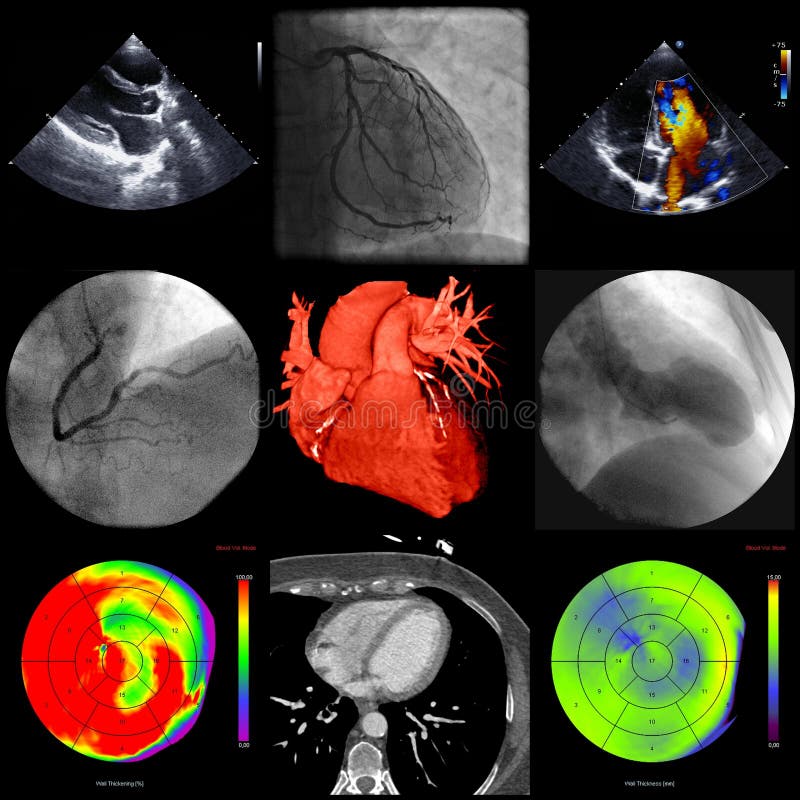
This technology uses the use of echoes from the waves produced to detect the structure and function of the heart. It includes transthoracic (TTE), transesophageal (TEE), and stress echocardiography.
Evaluation of heart failure (HF)
Valvular heart disease
Cardiomyopathy assessment
Pericardial effusion/tamponade
Infective endocarditis (TEE preferred)
Hemodynamic assessment in shock
Suspected structural heart disease
Ischemia evaluation (stress echo)
Real-time assessment of ventricular systolic and diastolic function
Valvular morphology and severity grading
Hemodynamic evaluation (e.g., pulmonary pressures)
Bedside utility in ICU/ED settings
No ionizing radiation
Widely available
Portable
Doppler allows flow quantification
First-line in heart failure
Operator-dependent
Image quality affected by lung disease
Limited tissue characterization compared to MRI
This technique provides real-time visuals of the cardiac function. It is the first-line imaging modality in assessing the heart morphology and pumping mechanism. It shows a clear view of the valvular anatomy and visualizes its abnormality.
Cardiac CT provides a clear visual of the intricate soft tissue components of the heart by providing cross-sectional views of selected segments. It provides high-resolution anatomical imaging, especially of the coronary arteries. It includes coronary CT angiography (CCTA) and calcium scoring.
Evaluation of stable chest pain with low–intermediate pre-test probability of CAD
Coronary artery anomaly assessment
Pre-procedural planning (TAVR, pulmonary vein isolation)
Aortic pathology (e.g., dissection)
Congenital heart disease
Assessment of bypass grafts
Excellent negative predictive value for obstructive CAD
Rapid acquisition
Noninvasive alternative to invasive coronary angiography
High spatial resolution
Coronary artery visualization
Analysis of coronary artery calcium (CAC score)
Useful in acute chest pain protocols
Ionizing radiation
Iodinated contrast exposure
Limited functional assessment
Overestimation of stenosis in heavily calcified vessels
It is preferred in cases of stable heart diseases rather than hemodynamics or myocardial tissue identification.
Provides high-resolution structural, functional, and tissue characterization without any ionizing radiation.
Cardiomyopathy characterization (e.g., HCM, DCM, ARVC)
Myocarditis evaluation
Viability assessment post-MI
Infiltrative diseases
Congenital heart disease
Cardiac masses
Iron overload assessment
Gold standard for ventricular volumes and ejection fraction
Late gadolinium enhancement (LGE) identifies fibrosis/scar
Tissue mapping (T1/T2) for edema and infiltration
Superior reproducibility
Excellent tissue characterization
No ionizing radiation
Multiparametric imaging
High diagnostic accuracy in non-ischemic cardiomyopathies
Limited availability in some settings
Longer acquisition time
Contraindications (non-MRI-compatible devices, severe claustrophobia)
Gadolinium caution in advanced renal failure
Cardiac MRI is the reference standard for myocardial tissue characterization and cardiomyopathy workup. It is often used when echo findings are inconclusive or when detailed myocardial assessment is required.
First line → ECG
If STEMI → immediate reperfusion
If stable, low-intermediate risk → Cardiac CT
If unclear myocardial injury → MRI (e.g., MINOCA workup)
First-line → Echocardiography
If etiology unclear or suspected infiltrative disease → MRI
Low–intermediate probability → Cardiac CT
Known CAD or functional assessment → Stress imaging (echo, MRI)
ECG, echocardiography, cardiac CT, and cardiac MRI are not competing modalities but complementary tools in cardiovascular diagnostics.
ECG: Electrical assessment and acute triage
Echo: First-line structural and functional evaluation
Cardiac CT: Coronary anatomy and stable chest pain
Cardiac MRI: Tissue characterization and cardiomyopathy
Optimal utilization requires alignment with clinical presentation, diagnostic yield, patient comorbidities, and guideline recommendations. A multimodality approach, when indicated, enhances diagnostic precision and patient outcomes.
Various cardiac diagnostic tools are available to monitor and diagnose a variety of heart conditions. They are modern technologies that help us figure out the condition of the heart non-invasively. It is important to select the appropriate imaging modality based on clinical presentation, pre-test probability, hemodynamic status, and resource availability. Four cornerstone modalities are
Electrocardiography (ECG),
Echocardiography (Echo)
Cardiac computed tomography (CT), and
Cardiac magnetic resonance imaging (MRI)
Each technology works differently and may help identify specific cardiac structures and functions.
The ECG records the electrical impulses that travel through the cardiac muscle through surface electrodes that are placed at specific parts of the body. It shows a wave whose slight abnormality indicates specific cardiac disease. It is the universally used, cheap, and rapid method available.
Suspected acute coronary syndrome (ACS)
Arrhythmias (AF, SVT, VT, bradyarrhythmias)
Conduction disorders (AV block, bundle branch block)
Electrolyte abnormalities (e.g., hyperkalemia)
Drug toxicity (e.g., QT prolongation)
Pericarditis
Initial evaluation of syncope, chest pain, or dyspnea
First-line diagnostic tool in emergency and outpatient settings
Identifies ST-segment elevation requiring emergent reperfusion
Provides rhythm diagnosis within a short duration
Essential for monitoring therapy and device function
Immediate results
No radiation
Bedside accessibility
Cost-effective
ECGs are a first-line diagnostic tool that only measure the electrical conductivity and not the structural and functional aspects. It could only be used as an adjunct for diagnosis. Utilizing other imaging modalities is required to arrive at the final diagnosis.
This technology uses the use of echoes from the waves produced to detect the structure and function of the heart. It includes transthoracic (TTE), transesophageal (TEE), and stress echocardiography.
Evaluation of heart failure (HF)
Valvular heart disease
Cardiomyopathy assessment
Pericardial effusion/tamponade
Infective endocarditis (TEE preferred)
Hemodynamic assessment in shock
Suspected structural heart disease
Ischemia evaluation (stress echo)
Real-time assessment of ventricular systolic and diastolic function
Valvular morphology and severity grading
Hemodynamic evaluation (e.g., pulmonary pressures)
Bedside utility in ICU/ED settings
No ionizing radiation
Widely available
Portable
Doppler allows flow quantification
First-line in heart failure
Operator-dependent
Image quality affected by lung disease
Limited tissue characterization compared to MRI
This technique provides real-time visuals of the cardiac function. It is the first-line imaging modality in assessing the heart morphology and pumping mechanism. It shows a clear view of the valvular anatomy and visualizes its abnormality.
Cardiac CT provides a clear visual of the intricate soft tissue components of the heart by providing cross-sectional views of selected segments. It provides high-resolution anatomical imaging, especially of the coronary arteries. It includes coronary CT angiography (CCTA) and calcium scoring.
Evaluation of stable chest pain with low–intermediate pre-test probability of CAD
Coronary artery anomaly assessment
Pre-procedural planning (TAVR, pulmonary vein isolation)
Aortic pathology (e.g., dissection)
Congenital heart disease
Assessment of bypass grafts
Excellent negative predictive value for obstructive CAD
Rapid acquisition
Noninvasive alternative to invasive coronary angiography
High spatial resolution
Coronary artery visualization
Analysis of coronary artery calcium (CAC score)
Useful in acute chest pain protocols
Ionizing radiation
Iodinated contrast exposure
Limited functional assessment
Overestimation of stenosis in heavily calcified vessels
It is preferred in cases of stable heart diseases rather than hemodynamics or myocardial tissue identification.
Provides high-resolution structural, functional, and tissue characterization without any ionizing radiation.
Cardiomyopathy characterization (e.g., HCM, DCM, ARVC)
Myocarditis evaluation
Viability assessment post-MI
Infiltrative diseases
Congenital heart disease
Cardiac masses
Iron overload assessment
Gold standard for ventricular volumes and ejection fraction
Late gadolinium enhancement (LGE) identifies fibrosis/scar
Tissue mapping (T1/T2) for edema and infiltration
Superior reproducibility
Excellent tissue characterization
No ionizing radiation
Multiparametric imaging
High diagnostic accuracy in non-ischemic cardiomyopathies
Limited availability in some settings
Longer acquisition time
Contraindications (non-MRI-compatible devices, severe claustrophobia)
Gadolinium caution in advanced renal failure
Cardiac MRI is the reference standard for myocardial tissue characterization and cardiomyopathy workup. It is often used when echo findings are inconclusive or when detailed myocardial assessment is required.
First line → ECG
If STEMI → immediate reperfusion
If stable, low-intermediate risk → Cardiac CT
If unclear myocardial injury → MRI (e.g., MINOCA workup)
First-line → Echocardiography
If etiology unclear or suspected infiltrative disease → MRI
Low–intermediate probability → Cardiac CT
Known CAD or functional assessment → Stress imaging (echo, MRI)
ECG, echocardiography, cardiac CT, and cardiac MRI are not competing modalities but complementary tools in cardiovascular diagnostics.
ECG: Electrical assessment and acute triage
Echo: First-line structural and functional evaluation
Cardiac CT: Coronary anatomy and stable chest pain
Cardiac MRI: Tissue characterization and cardiomyopathy
Optimal utilization requires alignment with clinical presentation, diagnostic yield, patient comorbidities, and guideline recommendations. A multimodality approach, when indicated, enhances diagnostic precision and patient outcomes.
Explore Topics
0%
Explore Topics
0%

